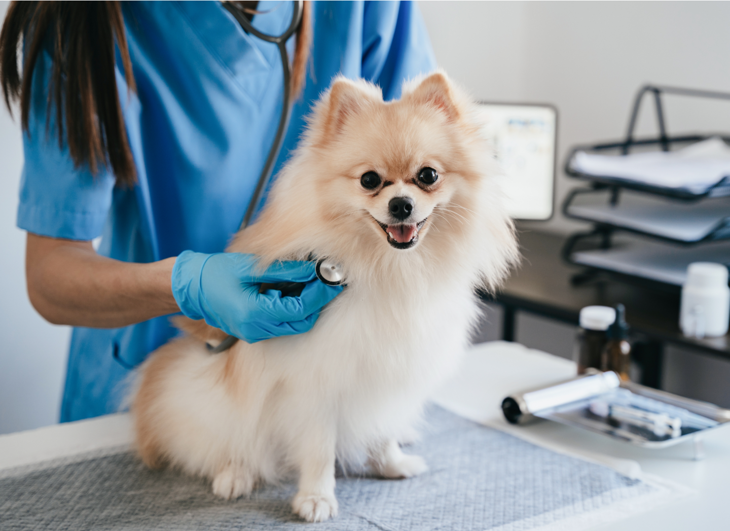
ZOETIS DIAGNOSTICS


Comprehensive urine sediment analysis
Powerful urine sediment analysis enabled by deep-learning AI provides consistent, accurate results within minutes1,2 for informed diagnosis and rapid treatment decisions.
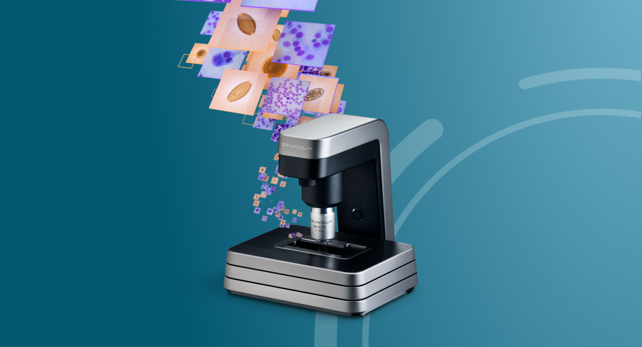
Urine sediment analysis is one of seven tests available with Vetscan Imagyst®, the world’s most capable veterinary AI analyzer.*
Help improve patient outcomes with convenient urine sediment analysis
- AI-powered analysis for results within minutes1,2
- Evaluates ~1000 fields of view for critical urine sediment elements
- Wide range of high-value diagnostic results with fresh samples3
- Fast, simple setup for maximum efficiency
- Easy slide preparation for consistent results
- Receive specialist support for diagnosis and treatment decisions when you need it†
Part of the complete, in-clinic urinalysis solution
Efficient, reliable data
AI-driven analysis with high performance accuracy comparable to that of a clinical pathologist guides diagnosis and treatment selection.1,2
Optimal workflow
Fast and simple slide preparation with consistent results allows you and your staff to complete testing and provide informative diagnostic insights in a single visit.
Advanced patient care
Part of a connected suite of urinalysis diagnostic tools, AI Urine Sediment maximizes your diagnostic capacity so you can provide continuous care with fewer delays to more patients.
How it works
- Highly accurate1,2 urine sediment analysis in just a few simple steps
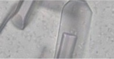
- Identifies red blood cells, white blood cells, epithelial cells, hyaline and non-hyaline casts, cocci and rod bacteria, spermatozoa and the following crystal types: struvite, calcium oxalate dihydrate, bilirubin, cystine and ammonium biurate
- Aids in the diagnosis of urinary tract pathology
Reliable urinalysis insights at the point of care
The proof is in the data — explore the diagnostic power of the Vetscan Imagyst® AI Urine Sediment application. See how AI-powered detection of urine sediment elements compares to that of expert, board-certified veterinary clinical pathologists.1,2,4
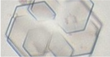
Familiar, easy-to-interpret images
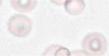
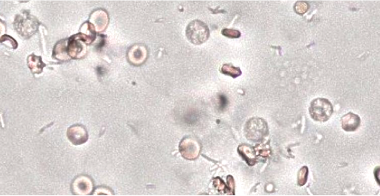
Red Blood Cells
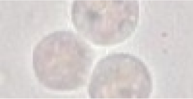
White Blood Cells
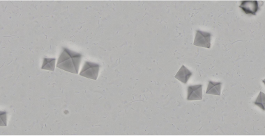
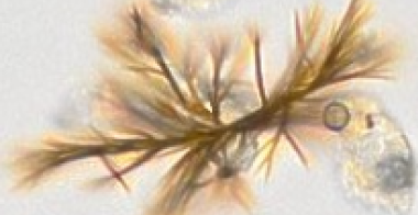
Calcium Oxalate Dihydrate Crystals
Struvite Crystals
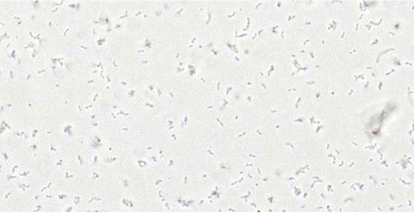
Cocci Bacteria
Rod Bacteria
Bilirubin Crystals
Cystine Crystals
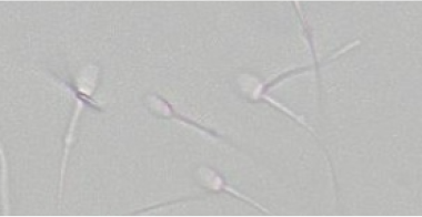
Spermatazoa
Frequently Asked Questions
The Vetscan Imagyst AI Urine Sediment application is verified for canine and feline urine sediment evaluations. The Vetscan Imagyst AI Urine Sediment algorithm will locate and quantify:
- Red Blood Cells (RBCs)
- White Blood Cells (WBCs)
- Squamous and Other (Renal tubular and Urothelial) Epithelial Cells
- Struvite Crystals
- Calcium Oxalate Dihydrate Crystals
- Bilirubin Crystals
- Cystine Crystals
- Ammonium Biurate Crystals
- Hyaline Casts
- Non-hyaline Casts
- Cocci bacteria
- Rod bacteria
- Spermatozoa
Source:
Data on file, Study No. DHXMZ-US-24-275, 2024, Zoetis Inc.
Data on file, Study No. DHXMZ-US-24-276, 2024, Zoetis Inc.
- Vetscan Imagyst urine sample prep device (consisting of urine centrifugation tube with graduated 1-, 2- and 3-mL fill lines and pipette tip with stopper)*
- Centrifuge capable of 450-500 RCF X 2 min
- Micropipette capable of 20μL sample (included with Vetscan Imagyst Welcome Kit)
- Preprinted slides with circular fiducial and frosted ends*
- Pencil to label patient ID on frosted end
- 22 X 22 mm coverslip*
* Included in the AI Urine Sediment Kit.
The sample preparation workflow is very similar, but there are a few very important differences. Both traditional and Vetscan Imagyst sample preparation involve centrifugation of the urine sample, pouring off supernatant, resuspending remaining pellet and placing a drop of urine sediment on a slide.
There are two main differences:
1. Standardization of sample preparation and evaluation: There is often little standardization with the traditional urine sediment sample preparation and evaluation. With the Vetscan Imagyst sample preparation, starting volume, residual volume and the amount of urine sediment placed on the slide will all be standardized. In addition, the sample will be evaluated in exactly the same way, every single time, by an algorithm that does not get tired at the end of the day.
2. The technician is able to perform other tasks while the Vetscan Imagyst AI Urine Sediment application is evaluating the sample.
Source:
Data on File. Study Report No. DHXMZ-US-23-218, 2023, Zoetis Inc.
Downloadable Resources
Looking for more information? Find what you need in our Resource Center.


Bringing specialist-level medicine to your clinic
The Zoetis Virtual Laboratory is an integrated support network of board-certified specialists paired with expert-level AI1,2,5-14, providing actionable insights to help you diagnose and treat with confidence.
- Best-in-class AI
- Anytime‡ expert support
- Connected diagnostic insights
Explore the full Vetscan Imagyst portfolio
Small, mighty and picture perfect, Vetscan Imagyst delivers insights across seven powerful testing capabilities.
* Vetscan Imagyst is the only commercially available veterinary AI analyzer on the market offering seven microscopic testing capabilities.
† Option to send digital slide image to our network of clinical pathologists as needed. Additional costs may apply.
‡ Dependent on consultant availability.
§ If you are a pet owner looking for treatment recommendations, please contact your veterinarian.
References: 1. Data on file, Study No. DHX6Z-US-24-275, 2024, Zoetis Inc. 2. Data on file, Study No. DHX6Z-US-24-276, 2024, Zoetis Inc. 3. Skeldon, N, et al. BSAVA Manual of Canine and Feline Clinical Pathology, 3rd Edition. Quedgeley, England: British small Animal Veterinary Association; 2016. pg. 184. 4. Data on file, Study No. DHXZ-US-23-218, 2024, Zoetis Inc. 5. Data on file, Study No. DHX6Z-US-23-205, 2024, Zoetis Inc. 6. Data on file, Study No. DHX6Z-US-23-206, 2024, Zoetis Inc. 7. Data on file, Study No. DHX6Z-US-23-209, 2024, Zoetis Inc. 8. Data on file, Study No. DHX6Z-US-24-257, 2024, Zoetis Inc. 9. Data on file. Study No. DHX6Z-US-24-242, 2024, Zoetis Inc. 10. Data on file, Study No. DHX6Z-US-23-222, 2023, Zoetis Inc. 11. Data on file, Study No. DHX6Z-US-22-131, 2022, Zoetis Inc. 12. Data on file, Study No. DHXMZ-US-25-285, 2025, Zoetis Inc. 13. Data on file, Study No. DHXMZ-US-25-286, 2025, Zoetis Inc. 14. Data on file. Study No. DHXMZ-US-24-235, 2024, Zoetis Inc.