ZOETIS DIAGNOSTICS


The world’s most capable veterinary AI analyzer*
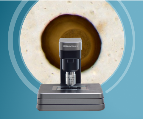
Transform your clinic with seven-in-one diagnostics from Vetscan Imagyst®, offering highly accurate sample analysis1-11 and high-definition imaging for deeply informed insights.
One analyzer, seven tests, endless insights
- Expert-level insights1-11 across AI-powered capabilities
- Remote support from a global network of clinical pathologists and parasitologists†
- Convenient access to digital cytology results with flexible delivery options
- AI Fecal, AI Urine Sediment, AI Dermatology, AI Blood Smear, AI Equine Fecal Egg Count, AI Masses and Digital Cytology included
- Easy-to-interpret images that reflect how cells and objects appear microscopically
- Small footprint with a sleek, compact design
-
Dimensions:7.09 x 7.09 x 7.48 in, 18 x 18 x 19 cm (HxWxD)
-
Weight:3.5 kg (7.72 lb)
-
Power:Use only power supply provided with scanner (100-240 VAC 50-60 Hz); use BBU if available
-
Connectivity:1 GigE wired connection; 802.11 ac WLAN
-
Operating Temperature:15-40° C (59-104° F)
-
Operating humidity:0-95% RH, non condensing
-
Operating altitude:Max. 2000 m
-
Storage temperature:1-70° C (34-158° F)
-
Slide capacity:1
-
Numerical aperture:0.75
-
Resolution, microscope:40X 0.25 μm/pix
First-of-its-kind innovation that keeps getting better
Small, mighty and picture perfect, Vetscan Imagyst delivers point-of-care insights, including AI-powered applications with expert-level results in minutes.1-9
AI Fecal
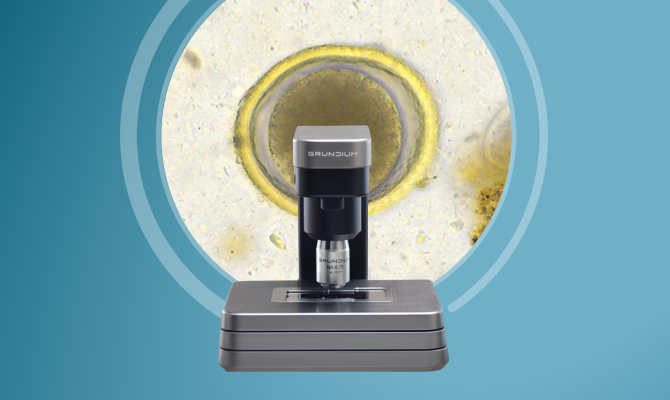
Clean, efficient approach to fecal analysis that accurately detects parasite ova, cysts and oocysts with results in minutes.4,5
AI Urine Sediment
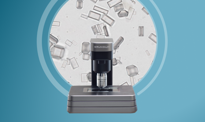
Consistent, accurate6,7 urine sediment analysis at the point of care that significantly reduces sample degradation concerns.
AI Dermatology
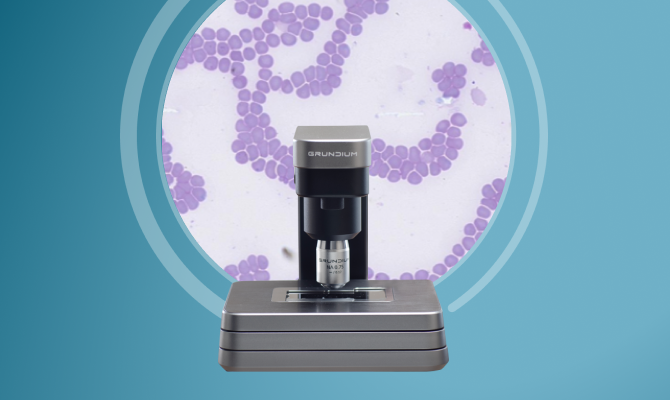
Powerful dermatology diagnostics that identify yeast, inflammatory cells and bacteria and differentiate between cocci and rods.8
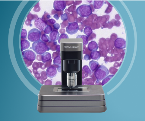
AI Blood Smear
Highly accurate blood smear qualitative analysis to supplement CBC results for a comprehensive hematology picture in minutes.1-3
AI Equine Fecal Egg Count
In-house equine FEC analysis with fast, simple sample preparation for reliable differentiation and quantification of Strongyles and Parascaris spp.9
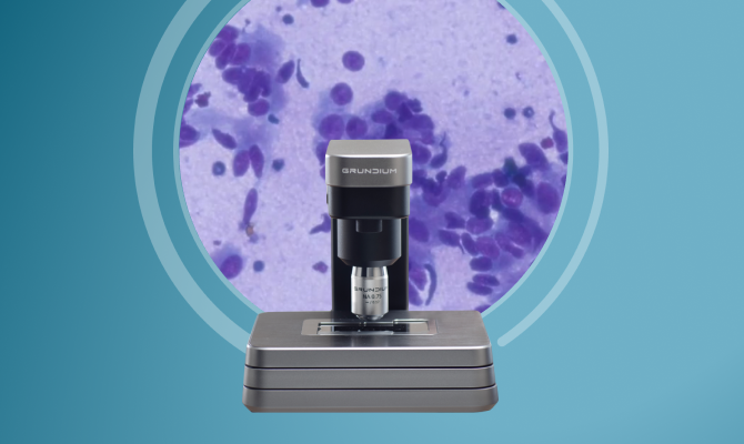
AI MassesNEW
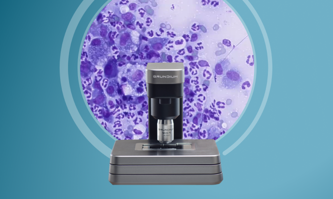
Cutting-edge AI analysis of common lymph node and skin/subcutaneous masses, completing our comprehensive in-clinic-cytology offering.10,11
Digital Cytology
Digital access to board-certified clinical pathologists that transforms the way you get cytopathology results with specialist insights within hours.‡,12
Frequently Asked Questions
Vetscan Imagyst is a first-of-its kind, multiuse platform that enables real-time, expert medical services and results through an AI and digital microscopy solution. It is the only in-clinic diagnostic platform with seven capabilities: AI Fecal, AI Urine Sediment, AI Dermatology, AI Blood Smear, AI Equine Fecal Egg Count, AI Masses and Digital Cytology image transfer capabilities. Vetscan Imagyst integrates image recognition technology with advanced AI and digital access to expert clinical pathologists within the Zoetis network to help you provide the best possible care. With a small in-office digital scanner, Vetscan Imagyst delivers accurate results comparable to a clinical pathologist or diagnostic parasitology expert.
Sources:
Data on file, Study No. DHX6Z-US-23-205, 2024, Zoetis Inc.
Data on file, Study No. DHX6Z-US-23-206, 2024, Zoetis Inc.
Data on file, Study No. DHX6Z-US-23-209, 2024, Zoetis Inc.
Data on file, Study No. DHX6Z-US-24-257, 2024, Zoetis Inc.
Data on file, Study No. DHX6Z-US-24-242, 2024, Zoetis Inc.
Data on file, Study No. DHX6Z-US-24-275, 2024, Zoetis Inc.
Data on file, Study No. DHX6Z-US-24-276, 2024, Zoetis Inc.
Data on file, Study No. DHX6Z-US-23-222, 2023, Zoetis Inc.
Data on file, Study No. DHX6Z-US-22-131, 2022, Zoetis Inc.
Data on file, Study No. DHXMZ-US-25-285, 2025, Zoetis Inc.
Data on file, Study No. DHXMZ-US-25-286, 2025, Zoetis Inc.
Vetscan Imagyst is a single system with multiple applications that uniquely combines expert diagnostics with AI capabilities, delivering efficient, actionable results.
The Vetscan Imagyst AI algorithms offer “set it and forget it” simplicity. It allows busy technicians and veterinarians to spend their valuable time examining pets and educating pet owners rather than evaluating samples under a microscope.
The Vetscan Imagyst system can also bring value to customers. Pet owners can see the images on the final report, adding a perception of value not typically found with current testing modalities. The images can make the results more tangible and provide an opportunity for excellent communication between you and your client. This may, in turn, lead to improved compliance by pet owners.
In addition, all Vetscan Imagyst customers have access to Virtual Laboratory by Zoetis for complimentary consultations with our global network of clinical specialists. Want to discuss results or a complex case? Simply book an appointment through ZoetisDx to connect via Zoom or email.*
* The use of Vetscan® Fuse/ Vetscan Hub™ plus at least one Zoetis Diagnostics instrument or service, such as Vetscan Imagyst, required.
Zoom is a registered trademark of Zoom Video Communications, Inc.
Currently, AI Fecal, AI Blood Smear, AI Dermatology, AI Urine Sediment and AI Masses are validated for canine and feline samples. AI Equine Fecal Egg Count is validated for equine fecal samples. The Digital Cytology application is available for all non-human species.
Yes, all AI algorithms are trained by boarded specialists and then subject to rigorous validation studies prior to being available for commercial use. This is true for AI Fecal, AI Urine Sediment, AI Dermatology, AI Blood Smear, AI Equine Fecal Egg Count, and AI Masses. In general, samples were prepared and scanned with the digital scanner. The expert read the glass slide and/or the digital whole slide image. Then the algorithm evaluated the sample, and the results were compared. In addition, for AI Fecal and AI Urine Sediment, the sample preparation devices were validated against a standard reference laboratory sample preparation method.
Sources:
Data on file, Study No. DHX6Z-US-23-205, 2024, Zoetis Inc.
Data on file, Study No. DHX6Z-US-23-206, 2024, Zoetis Inc.
Data on file, Study No. DHX6Z-US-23-209, 2024, Zoetis Inc.
Data on file, Study No. DHX6Z-US-24-257, 2024, Zoetis Inc.
Data on file, Study No. DHX6Z-US-24-242, 2024, Zoetis Inc.
Data on file, Study No. DHX6Z-US-24-275, 2024, Zoetis Inc.
Data on file, Study No. DHX6Z-US-24-276, 2024, Zoetis Inc.
Data on file, Study No. DHX6Z-US-23-222, 2023, Zoetis Inc.
Data on file, Study No. DHX6Z-US-22-131, 2022, Zoetis Inc.
Data on file, Study No. DHXMZ-US-25-285, 2025, Zoetis Inc.
Data on file, Study No. DHXMZ-US-25-286, 2025, Zoetis Inc.
WSI refers to the scanning of glass microscope slides to produce digital images. Vetscan Imagyst uses the Grundium Ocus®40 slide scanner to capture high-resolution images of the entire slide. The upload of the image to the cloud enables an expert clinical pathologist or parasitologist to examine the specimen as if they were looking at the slide through a microscope at the veterinary clinic. AI Dermatology, AI Blood Smear, AI Masses and Digital Cytology image transfer scan one layer or depth on the slide. AI Fecal scans 2 distinct layers where the parasite eggs, cysts and oocysts are expected to be floating, and AI Urine Sediment scans 5 different layers to capture all of the common urine sediment elements. This means the WSI for AI Urine Sediment consists of 5 different layers of the whole slide.
Ocus is a registered trademark of Grundium Oy
The Vetscan Imagyst AI Dermatology application is an efficient and accurate way to perform in-clinic dermatologic cytology analysis within minutes–giving you more time with patients. The Imagyst can aid in the detection and diagnosis of skin infections through use of advanced AI technology and deliver consistent results, regardless of personnel, training or microscope performance. The AI Dermatology application reviews impression smears, ear swabs and skin swabs to detect the presence of yeast, inflammatory cells and bacteria as well as differentiate between cocci and rods. Application users also have the option to have their cytology image submitted for expert review by our network of board certified pathologists through Virtual Laboratory by Zoetis (additional costs may apply).
With the AI Blood Smear application, simply prepare a slide using traditional methods, then scan using the Vetscan Imagyst digital scanner and allow AI to perform the analysis. Vetscan Imagyst uses AI-driven analysis to:
- Clarify abnormalities to supplement automated complete blood count (CBC) results
- Verify a white blood cell (WBC) differential (%) and estimated relative count of each cell type
- Included in WBC differential is band (immature) neutrophils
- Provide an estimated platelet count and identify the presence of platelet clumps, which may impact platelet counts
- Identify and count polychromatophils (immature red blood cells—an indicator of a potential regenerative process) and nucleated red blood cells (nRBC)
- Identify the presence of poikilocytes (acanthocytes, echinocytes, keratocytes, schistocytes, eccentrocytes)
AI Fecal analysis for Vetscan Imagyst was developed to provide a simple, easy and systematized fecal examination that is less influenced by different fecal preparation methods or the skill level of an examiner. By leveraging deep learning, the fecal application uses cutting-edge image recognition technology to help improve fecal in-clinic diagnostics by reducing user variability and human error. Vetscan Imagyst AI Fecal can also help expedite treatment intervention and will maintain shareable records for future needs. Three peer-reviewed studies published between Parasites & Vectors and JVDI demonstrated that Vetscan Imagyst AI Fecal analysis performed well versus a diagnostic parasitology expert evaluating the same sample with expert focus and attention—something that is less likely to be done with each in-office fecal exam.
The digital cytology application is a fast, convenient approach to expert clinical pathology. With efficient review of common cytology specimens, such as fine-needle aspirates, Vetscan Imagyst helps veterinarians provide the best possible care. Vetscan Imagyst accelerates the process for expert clinical pathology review—simply prepare and scan slides and then submit for expert results in hours. Submissions are prepared in clinic using traditional methods, but submitted digitally, rather than sending physical slides to a laboratory.
The AI Urine Sediment application delivers fast, efficient urine sediment analysis through the digital scanning and AI evaluation of a urine sediment wet prep sample. The Vetscan Imagyst Urine Sediment sample preparation method has been created to standardize processes and results, no matter who is preparing the sample. With algorithm accuracy comparable to that of board-certified clinical pathologists, users can expect consistent, reliable results for each analysis regardless of personnel, training or microscopic performance.
Vetscan Imagyst AI Equine Fecal Egg Count analysis has an easy sample preparation process that is very similar to the steps of the widely used Modified McMaster Technique, but rather than being counted in a chamber, utilizes a concentration technique for egg collection. Vetscan Imagyst AI Equine Fecal Egg Count analysis is powered by deep learning image recognition technology to help veterinarians make accurate and timely intestinal parasite diagnoses for patients at the point of care and complement the work of a veterinary professional. Fecal Egg Count (FEC) results are provided in eggs per gram (EPG), to identify low, medium, and high shedders as described by the American Association of Equine Practitioners (AAEP) Internal Parasite Control Guidelines. Results from verification studies showed that the algorithm performed comparable to a diagnostic parasitology expert and provided expert level results in about 7 minutes.
Vetscan Imagyst AI Masses enables you to rapidly identify cells suggestive of pathology in common lymph node and skin/subcutaneous masses at the point of care, powered by deep-learning AI and supported by add-on remote expert clinical pathology review when clinically warranted.* Slide preparation for AI Masses follows the same procedures as Digital Cytology: Prepare the sample using a fine needle aspirate (FNA) or fine needle biopsy (FNB) technique, following industry best practices.
The digital cytology application is a fast, convenient approach to expert clinical pathology. With efficient review of common cytology specimens, such as fine-needle aspirates, Vetscan Imagyst helps veterinarians provide the best possible care. Vetscan Imagyst accelerates the process for expert clinical pathology review—simply prepare and scan slides and then submit for expert results delivered when you need them.‡. Submissions are prepared in clinic using traditional methods, but submitted digitally, rather than sending physical slides to a laboratory.
With a small in-office footprint, Vetscan Imagyst delivers a streamlined approach to fecal, urine sediment, dermatology, blood smear, equine fecal egg count, and cytology testing. As new applications become available, they will be easily integrated with existing Vetscan Imagyst testing capabilities.
*Additional costs may apply.
Sources:
Data on file, Study No. DHX6Z-US-23-222, 2023, Zoetis Inc.
Data on file, Study No. DHX6Z-US-23-205, 2024, Zoetis Inc.
Data on file, Study No. DHX6Z-US-23-206, 2024, Zoetis Inc.
Data on file, Study No. DHX6Z-US-23-209, 2024, Zoetis Inc.
Data on file, Study No. DHX6Z-US-24-257, 2024, Zoetis Inc.
Data on file, Study No. DHX6Z-US-24-242, 2024, Zoetis Inc.
Data on file, Study No. DHXMZ-US-24-275, 2024, Zoetis Inc.
Data on file, Study No. DHXMZ-US-24-276, 2024, Zoetis Inc.
Data on file. Study No. TI-11711, 2024, Zoetis Inc.
Data on file, Study No. DHXMZ-US-23-218, 2023, Zoetis Inc.
Data on file, Study No. DHX6Z-US-22-131, 2022, Zoetis Inc.
American Association of Equine Practitioners. Internal Parasite Control Guidelines. Updated 2024.
Nagamori, Y., Hall Sedlak, R., DeRosa, A. et al. Evaluation of the VETSCAN IMAGYST: an in-clinic canine and feline fecal parasite detection system integrated with a deep learning algorithm. Parasites Vectors 13, 346 (2020).
Nagamori, Y., Sedlak, R.H., DeRosa, A. et al. Further evaluation and validation of the VETSCAN IMAGYST: in-clinic feline and canine fecal parasite detection system integrated with a deep learning algorithm. Parasites Vectors 14, 89 (2021).
Nagamori Y, Scimeca R, Hall-Sedlak R, et al. Multicenter evaluation of the Vetscan Imagyst system using Ocus 40 and EasyScan One scanners to detect gastrointestinal parasites in feces of dogs and cats. Journal of Veterinary Diagnostic Investigation. 2023;0(0). doi:10.1177/10406387231216185
Data on file, Study No. DHXMZ-US-25-285, 2025, Zoetis Inc.
Data on file, Study No. DHXMZ-US-25-286, 2025, Zoetis Inc.
Yes, if more information is needed in addition to the Vetscan Imagyst AI Urine Sediment, AI Dermatology or AI Blood Smear analyses, the scanned whole slide image (WSI) can be sent to a clinical pathologist using the Add-On Expert Review function.*
For AI Urine Sediment, if the Add-On Expert Review is requested, veterinarians are encouraged to submit an additional stained, air-dried urine sediment smear slide as part of the sample submission process. This will allow the clinical pathologists to provide more information, especially regarding cell morphology and bacteria.
These findings may provide important additional information that could guide diagnosis and/or treatment.
*Option to submit digital slide image to our network of clinical pathologists as needed. Additional costs may apply.
Not all image recognition AI is the same. The methods currently used in the veterinary market to train models and classify data are known as Superficial and Deep Learning. Two key differences between superficial and deep-learning algorithms are the way in which the algorithm is trained, and the number of “features” used in the classification of new objects. “Features” are distinguishing characteristics that visually make an object that object (e.g., a cat has whiskers, fur, two eyes, four legs, etc.). With superficial learning AI algorithms, a human manually selects the “features” of an object, and the algorithm uses these features to classify new objects.
In deep-learning AI algorithms, object “feature” selection and training are not limited by manual or human selection. This step is skipped. In deep learning, countless numbers of objects or data are shown to the algorithm, and the algorithm extracts relevant “features” that it uses to classify new objects. During the training process, an expert tells the algorithm whether it has classified objects correctly, allowing the AI to learn and extract more features with each correction. This process is illustrated in Figure 1 below.
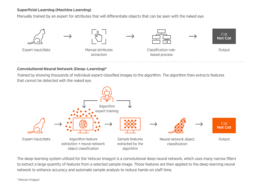
Figure 1. Types of Image Analysis AI Algorithms
In superficial learning fewer features are evaluated, because it relies on a human expert to identify every feature used to train the algorithm (i.e. the human tells the algorithm what attributes of a cell to pick out). It can be difficult to improve these algorithms as the human needs to visualize more features to teach the algorithm. On the other hand, deep-learning AI systems use thousands of features and their inter-relations to identify an object. The algorithm determines differentiating attributes that we often times cannot even pick up with our eyes (i.e. the human expert tells the algorithm, “this is a cell” and the algorithm breaks the image down to the pixel level to decide what key attributes define a “cell”). Improvements to deep-learning algorithms are generally related to providing more and better image examples so the combined feature extraction and analysis components of deep learning can be maximized. The deep-learning system that is utilized for the Vetscan Imagyst is a convolutional deep neural network. A convolutional deep neural network uses a very large number of small filters to extract a very large number of features from the image which are applied to the deep learning neural network. Deep-learning AI will excel in identification of often difficult to visualize elements compared to superficial learning AI.
The deep-learning nature of the Vetscan Imagyst algorithm allows for evolution and increased functionality over time. In the future, the algorithm may be trained to classify additional elements and further validation studies can be performed. Customer feedback is essential in helping to determine which elements may be trained into future iterations of the algorithms.
No. While expertly classified training and hold-out (model test) data is continuously added by our boarded specialists and algorithm team, this is tightly managed and not driven by the AI itself. Only algorithm updates that have been validated through studies are released to customers.
We are here to help! Contact us at DXsupport@Zoetis.com or 1-888-963-8471.
A high-speed internet connection is needed to best experience the benefits of Vetscan Imagyst.
Vetscan Imagyst is compatible with most practice information management software (PIMS) systems. Contact your Zoetis representative for the full list of compatible software.
The platform does not require a dedicated computer. In addition, only a small amount of benchtop space is required.
AI Fecal, AI Urine Sediment, AI Dermatology, AI Blood Smear, AI Equine Fecal Egg Count, and AI Masses all use AI to deliver results in minutes.
Digital Cytology Image Transfer: Vetscan Imagyst delivers fast digital access to clinical pathologists. For routine cases, choose Standard Review for next-day1 results. For more critical cases, Urgent Review can provide results in hours.1
Source:
Data on file. Study No. TI-11711, 2024, Zoetis Inc.
Downloadable Resources
Looking for more information? Find what you need in our Resource Center.


Bringing specialist-level medicine to your clinic
The Zoetis Virtual Laboratory is an integrated support network of board-certified specialists paired with expert-level AI1-11,13, providing actionable insights to help you diagnose and treat with confidence.
- Best-in-class AI
- Anytime§ expert support
- Connected diagnostic insights
Discover the difference Vetscan Imagyst can make in your clinic
Contact us today to learn more about how our diagnostics portfolio can help elevate your patient care.
Customer Service, Support & Ordering
Mon – Fri: 8:30am – 6:30pm ET
Vetscan & i-STAT Technical Support
24 hours a day, 7 days a week, 365 days a year
1-888-963-8471
Zoetis Reference Laboratories Customer Service, Support & Ordering
Mon – Fri: 8:00am – 9:30pm ET
Saturday: 9:00am – 8:30pm ET
Sunday: Closed
1-888-965-9652
Need Technical Support?
Our support team is available to answer product questions and provide guidance.§
* Vetscan Imagyst is the only commercially available veterinary AI analyzer on the market offering seven microscopic testing capabilities.
† Option to send digital slide image to our network of clinical parasitologists or pathologists as needed. Additional costs may apply.
Complimentary AI Fecal image review from a support specialist is available for concerning results.
‡ Additional costs may apply
§ Dependent on consultant availability.
|| If you are a pet owner looking for treatment recommendations, please contact your veterinarian.
References: 1. Data on file, Study No. DHX6Z-US-23-205, 2024, Zoetis Inc. 2. Data on file, Study No. DHX6Z-US-23-206, 2024, Zoetis Inc. 3. Data on file, Study No. DHX6Z-US-23-209, 2024, Zoetis Inc. 4. Data on file, Study No. DHX6Z-US-24-257, 2024, Zoetis Inc. 5. Data on file. Study No. DHX6Z-US-24-242, 2024, Zoetis Inc. 6. Data on file, Study No. DHX6Z-US-24-275, 2024, Zoetis Inc. 7. Data on file, Study No. DHX6Z-US-24-276, 2024, Zoetis Inc. 8. Data on file, Study No. DHX6Z-US-23-222, 2023, Zoetis Inc. 9. Data on file, Study No. DHX6Z-US-22-131, 2022, Zoetis Inc. 10. Data on file, Study No. DHXMZ-US-25-285, 2025, Zoetis Inc. 11. Data on file, Study No. DHXMZ-US-25-286, 2025, Zoetis Inc. 12. Data on file, Study No. TI-11711, 2024, Zoetis Inc. 13. Data on file, Study No. DHXMZ-US-24-235, 2024, Zoetis Inc.