ZOETIS DIAGNOSTICS


Cytologic analysis at the point of care
Give pet owners the answers they need with fast, AI-powered analysis of common lymph node and subcutaneous masses, enabling accurate1,2 results for treatment and diagnosis on your timeline.
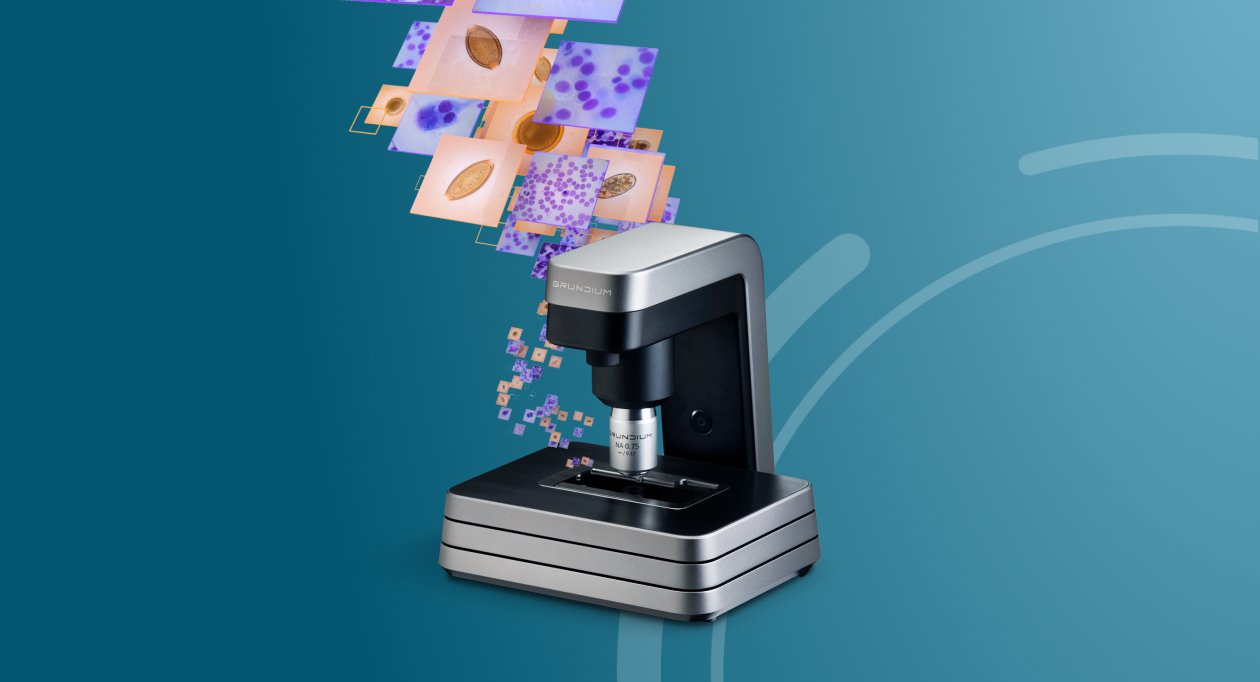
AI Masses is one of six tests available with Vetscan Imagyst®, the world’s most capable veterinary AI analyser.*
Reduce client anxiety with fast, in-clinic cytologic analysis
- AI powered analysis for results within minutes1,2
- Evaluates potentially cancerous cells in common lymph node and subcutaneous masses
- Clear, detailed images for informed decisions
- Familiar slide preparation without changing your workflow
- Expert clinical pathologist review when clinically warranted†
- In-clinic screening for more accessible testing
Part of the complete, in-clinic cytologic solution
Efficient, reliable results
In-clinic screening of potentially cancerous cells in common lymph node and subcutaneous masses with best-in-class AI enables fast, accurate1,2 diagnosis and treatment.
Optimal workflow
Familiar, one-time sample preparation and rapid results save time while allowing your clinic to maintain your current workflow.
Advanced patient care
Help improve compliance and reduce pet owner stress by providing accessible, in-clinic screening.
How it works
- Highly accurate1,2 analysis of common lymph node and subcutaneous masses
- Identifies findings suggestive of large, medium, small or mixed lymphocyte population, inflammatory cells, plasma cells, reactive lymph node or neoplasia, histiocytoma, mast cell tumour, plasma cell tumour, large cell lymphoma, reactive lymphoid hyperplasia, lymph node with inflammation, not lymph node, lipoma/adipose tissue, keratin containing lesion or inflammation
- Fast, point-of-care screening unlocks individualised treatment for patients
Part of a comprehensive cytology offering
Unlock the freedom to make timely individualised diagnoses and treatment decisions that fit your patients’ needs with Vetscan Imagyst cytology.
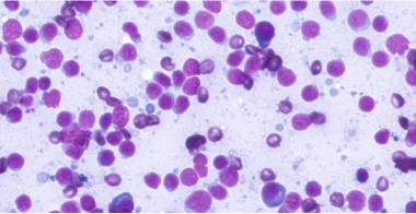
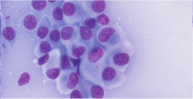
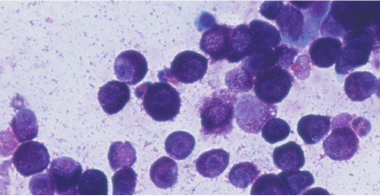
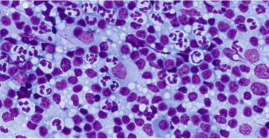
High-resolution images for more confident diagnoses
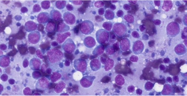
Large cell lymphoma (neoplasia)
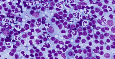
Lymph node with inflammation
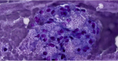
Adipose or salivary tissue
Reactive lymphoid hyperplasia
Histiocytoma
Mast cell tumour
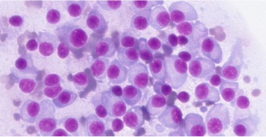
Plasma cell tumour
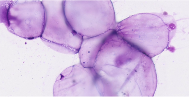
Lipoma/adipose tissue
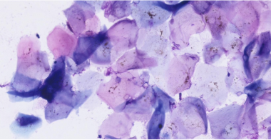
Keratin containing lesion
Inflammatory lesion
Downloadable Resources
Looking for more information? Find what you need in our Resource Centre


Frequently Asked Questions
Both Vetscan Imagyst AI Masses and Digital Cytology can help you provide quick answers to anxious pet owners. AI Masses allows you to test common lymph node and subcutaneous masses in-office for fast results in minutes. With Digital Cytology, you can send the samples to board-certified clinical pathologists for results delivered when you need them.‡,3
The types of clinical objects that can be identified via AI Masses include:
Lymph node:
- Large cell lymphoma (neoplasia)
- Lymph node with inflammation
- Not lymph tissue (adipose or salivary tissue)
- Reactive lymphoid hyperplasia
- Indeterminate results
Subcutaneous masses:
- Histiocytoma
- Mast cell tumor
- Plasma cell tumor
- Lipoma/adipose tissue
- Keratin containing lesion
- Inflammatory lesion
- Indeterminate results
As with all Vetscan Imagyst applications, you'll need a laptop, tablet, or mobile device and a Vetscan Imagyst scanner. In addition, you’ll need standard materials including 25 x 75 mm microscope glass slides with frosted edge, Romanowsky-type stain (e.g. Diff Quik), immersion oil, 24 x 60 mm coverslips, a pencil and gloves.
AI Masses allows you to test common lymph node and subcutaneous masses.
With the same familiar single-sample preparation, AI Masses doesn't require you to change your traditional workflow, allowing you to spend less time training staff and giving you more control over how your practice is run.

Bringing specialist-level medicine to your clinic
The Zoetis Virtual Laboratory is an integrated support network of board-certified specialists paired with expert-level AI1,2, 4-13, enhancing every element of your diagnostic practice to help you diagnose and treat with confidence.
- Best-in-class AI
- Anytime§ expert support
- Connected diagnostic insights
Explore the full Vetscan Imagyst portfolio
Small, mighty and picture perfect, Vetscan Imagyst delivers unmatched insights across six powerful testing capabilities.
Discover how Vetscan Imagyst can transform your practice
Contact us today to learn more about how our diagnostic portfolio can help you provide elevated patient care.
* Vetscan Imagyst is the only commercially available veterinary AI analyser on the market offering six microscopic testing capabilities.
† Option to send digital slide image to our network of clinical pathologists as needed. Additional costs may apply.
‡ Additional costs may apply
§ Dependent on consultant availability.
|| If you are a pet owner looking for treatment recommendations, please contact your vet.
References: 1.Data on file, Study No. DHX6Z-US-25-285, 2025, Zoetis Inc. 2. Data on file, Study No. DHX6Z-US-25-286, 2025, Zoetis Inc. 3. Data on file, Study No. TI-11711, 2024, Zoetis Inc. 4. Data on file, Study No. DHX6Z-US-23-205, 2024, Zoetis Inc. 5. Data on file, Study No. DHX6Z-US-23-206, 2024, Zoetis Inc. 6. Data on file, Study No. DHX6Z-US-23-209, 2024, Zoetis Inc. 7. Data on file, Study No. DHX6Z-US-24-257, 2024, Zoetis Inc. 8. Data on file. Study No. DHX6Z-US-24-242, 2024, Zoetis Inc. 9. Data on file, Study No. DHX6Z-US-24-275, 2024, Zoetis Inc. 10. Data on file, Study No. DHX6Z-US-24-276, 2024, Zoetis Inc. 11. Data on file, Study No. DHX6Z-US-23-222, 2023, Zoetis Inc. 12. Data on file, Study No. DHX6Z-US-22-131, 2022, Zoetis Inc. 13. Data on file. Study No. DHXMZ-US-24-235, 2024, Zoetis Inc.